The Clinical Necessity of Routine Ocular Evaluations
A basic vision screening tells you if you can read letters on a wall. A thorough ocular evaluation reveals the vascular and neurological health of your entire body. The distinction between the two dictates the level of care a patient receives. The decision to transition a patient from basic vision screenings to full exams is reached by evaluating systemic risk factors, such as a family history of diabetes or hypertension, rather than waiting for visual complaints. Waiting for symptoms often means waiting for irreversible damage.

Establishing a baseline is the cornerstone of optimal eye care. Based on longitudinal studies, baseline comparative analysis often requires initial retinal imaging to be captured between ages 35 and 40 for asymptomatic adults. This early documentation provides a structural map of the optic nerve and macula. When subtle changes occur a decade later, practitioners have a definitive reference point rather than relying on population averages.
Patient History and Preliminary Diagnostics
Gathering the Clinical Context
Every examination begins before the patient sits behind the diagnostic equipment. Collecting a detailed medical history, current medication list, and specific visual demands sets the trajectory for the entire visit. A patient working twelve hours a day on multiple monitors requires a different investigative approach than a retired commercial driver.
Tonometry and Corneal Curvature
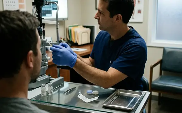
Preliminary diagnostics establish the physical parameters of the eye. Autorefraction and keratometry provide an immediate baseline of corneal curvature and estimated refractive error. Intraocular pressure measurement follows, presenting a classic clinical trade-off. Initially, non-contact tonometry was considered for all rapid screenings to minimize cross-contamination, but practitioners reverted to Goldmann applanation for patients with borderline high readings.
The reasoning is rooted in anatomical variance. Air-puff tonometry can overestimate intraocular pressure by 2 to 4 mmHg in patients with thicker-than-average corneas. Conversely, it can dangerously underestimate pressure in thinner corneas.
Risk Factor: Failure to detect early-stage normal-tension glaucoma in patients with naturally thin corneas when relying solely on non-contact tonometry.
Goldmann applanation tonometry remains the gold standard. It requires the instillation of a fluorescein dye and anesthetic drop, taking effect within 15 to 30 seconds. The physical contact with the cornea provides a highly accurate reading that dictates subsequent glaucoma management protocols.
Refraction and Visual Acuity Assessment
The Mechanics of Subjective Refraction
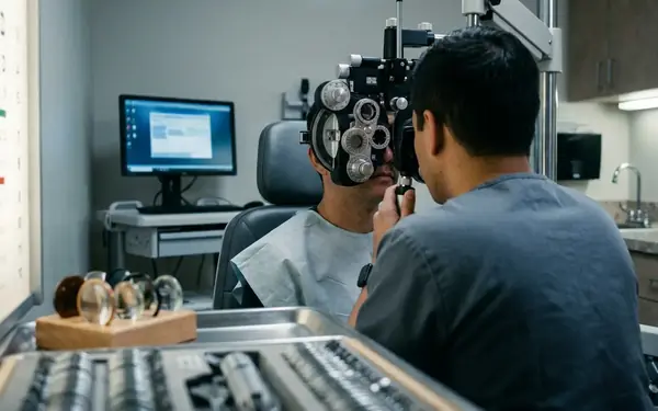
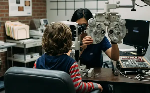
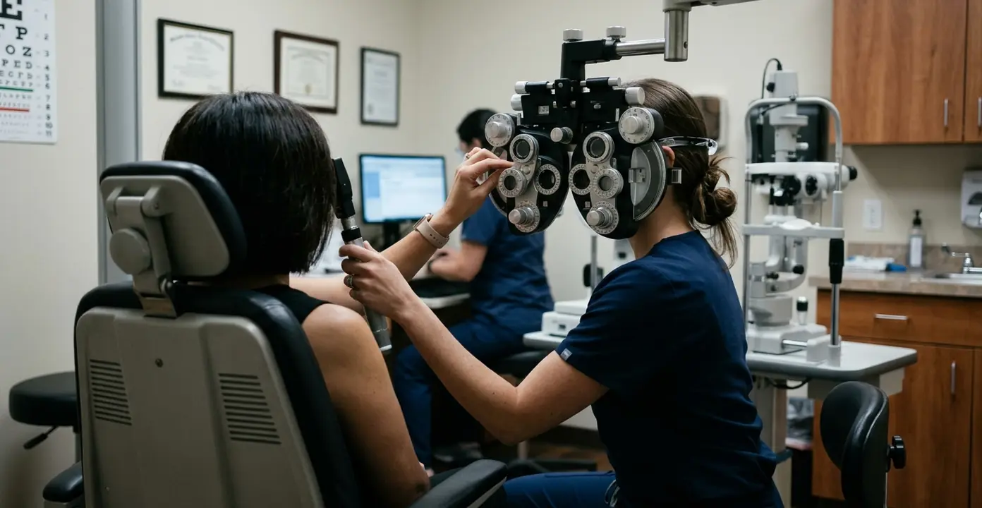
Determining a patient's precise prescription is a collaborative process. Standard subjective refraction typically isolates visual acuity using spherical and cylindrical lens increments of 0.25 diopters. The phoropter allows the practitioner to rapidly cycle through these proven lens combinations.
The endpoint of subjective refraction is determined by introducing a Jackson Cross Cylinder to isolate astigmatic axes, prompting the patient to compare clarity between two lens options until visual equality is achieved. This technique eliminates guesswork. It relies on the patient's direct perception of contrast and sharpness.
Binocular Vision Assessment
Clear vision in each eye individually does not guarantee comfortable vision in the real world. Assessment of binocular vision, eye teaming, and focusing capabilities ensures the eyes work as a synchronized unit. Deficits in convergence or accommodation often manifest as late-day headaches or reading fatigue, symptoms that a standard acuity check will completely miss.
Slit-Lamp Biomicroscopy and Retinal Evaluation
Anterior Segment Analysis
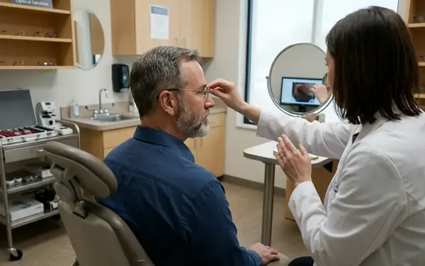
The slit-lamp biomicroscope provides a highly magnified, stereoscopic view of the anterior segment. We evaluate the cellular integrity of the cornea, the architecture of the iris, and the clarity of the crystalline lens. This microscopic examination identifies early cataract formation, dry eye disease markers, and anterior inflammation.
Pharmacological Dilation Protocols
Viewing the posterior segment requires expanding the pupil. The choice of pharmacological dilating agents involves assessing the patient's anterior chamber angle depth; a shallow angle prompts the use of a weaker mydriatic agent to prevent inducing acute angle-closure glaucoma. During protocol evaluations, standard presentations using Tropicamide 1% combined with phenylephrine at around 2.5% typically achieve maximum pupillary dilation within 20 to 35 minutes.
Once dilated, the evaluation of the optic nerve, macula, and peripheral retina begins. This is where systemic diseases often reveal their earliest ocular signs.
Recommendation: Patients should review dilated eye exam guidelines prior to their visit. Caveat: Pharmacological dilation induces photophobia and cycloplegia that impairs near-vision tasks for a duration of 4 to 6 hours post-instillation.
Diagnostic Limitations of Standard Examinations
The Boundaries of Direct Observation
Even with maximum dilation and expert technique, standard exams have physical boundaries. Standard binocular indirect ophthalmoscopy provides a field of view limited to approximately 30 to 40 degrees of the retina at any single moment. The practitioner must mentally stitch these views together to form a complete picture of the peripheral retina.
Furthermore, standard exams may not detect microscopic retinal layer anomalies. In clinical practice, the determination to order advanced Optical Coherence Tomography is made when slit-lamp biomicroscopy reveals subtle macular pigment changes that cannot be definitively classified as drusen or fluid accumulation. OCT provides cross-sectional imaging that resolves cellular layers down to the micron level.
Temporal Limitations
Standard exams represent a snapshot in time—a static view of a dynamic biological system. A normal pressure reading at 9:00 AM does not preclude dangerous pressure spikes at midnight. This temporal limitation underscores the necessity of adherence to customized follow-up schedules.
Post-Examination Protocols and Corrective Implementation
Synthesizing the Treatment Plan
Data collection concludes; clinical synthesis begins. Formulating a treatment plan requires integrating refractive data, binocular function, and ocular health findings. Prescribing optical interventions or medications is highly specific to the individual's daily environment. Customized optical interventions for progressive myopia often require an adaptation period reported as roughly 7 to 12 days for the visual cortex to adjust to new peripheral lens designs. Patients must be counseled on this transition to ensure compliance.
Establishing Recall Schedules
The final step is determining when the patient needs to return. Recall schedules are customized by synthesizing the severity of refractive shifts and retinal health; a stable myope is scheduled annually, whereas a patient exhibiting early microaneurysms is shifted to a 3-to-6-month interval. Recall schedule variations based on the presence of systemic autoimmune conditions requiring high-risk ocular medications are also strictly enforced.
Critical Insight: Through an ongoing partnership since 2019 with regional diabetic care centers, longitudinal data shows that strict adherence to customized recall intervals reduces the incidence of severe vision loss by catching proliferative changes before they become symptomatic.
Patient Preparation Checklist for Full Eye Exams
- Compile a list of current systemic medications, particularly corticosteroids or immunosuppressants.
- Bring current prescription eyewear, including backup spectacles and contact lens packaging.
- Arrange transportation if pharmacological dilation is planned.
No single diagnostic snapshot is guaranteed to predict long-term progression without consistent follow-up. While full exams detect most structural anomalies, functional vision fluctuations require ongoing patient reporting. Protecting your sight is a continuous, collaborative effort between patient and practitioner.