The Pathophysiology of Glaucoma
Glaucoma is rarely a single, isolated disease. It is a group of progressive optic neuropathies characterized by the gradual deterioration of the optic nerve. Understanding this condition requires looking at the fluid dynamics within the anterior chamber of the eye.
The healthy ciliary body produces aqueous humor at about 2 to 3 microliters per minute. This clear fluid nourishes the avascular structures of the eye before draining through the trabecular meshwork. Across multi-center studies, normal intraocular pressure is traditionally defined as resting between 10 and 21 mmHg. When the drainage system encounters resistance, fluid accumulates, and pressure inside the eye begins to rise.
Historically, elevated pressure was considered the sole culprit. However, clinicians shifted to a dual-mechanism model—incorporating both mechanical compression and vascular insufficiency, after observing progressive retinal ganglion cell death in patients whose intraocular pressure was strictly controlled. Mechanical stress physically damages the nerve fibers at the lamina cribrosa, while vascular insufficiency starves those same fibers of essential oxygen and nutrients.
Primary and Secondary Risk Factors
Demographic variables establish the baseline risk for developing glaucomatous damage. Age over 60 and a direct family history of the disease are well-documented primary indicators. Epidemiological assessments indicate that individuals of African descent face an onset of primary open-angle glaucoma roughly 10 to 15 years earlier than Caucasian cohorts.
Systemic health plays an equally critical role. Through an ongoing clinical partnership since 2019 with regional endocrinology centers, assessment confirmed that patients with hypertension, diabetes, and cardiovascular disease face compounded risks. These conditions compromise the delicate microvasculature supplying the optic nerve head.
Ocular anatomy provides the final piece of the risk profile. High axial myopia stretches the ocular tissues, making the optic nerve more susceptible to damage. Corneal thickness is another vital metric. Ophthalmologists adjust baseline risk profiles by cross-referencing intraocular pressure with pachymetry readings, deciding to escalate monitoring frequency when central corneal thickness measures below average.
Risk Factor: From screening data, central corneal thickness measuring below 500 micrometers significantly increases the risk of underestimating true intraocular pressure.
Asymptomatic Progression and Symptom Onset
Primary open-angle glaucoma is universally termed the 'silent thief of sight' because it operates without early warning signs. In my clinical practice, I frequently see patients who assume a passed vision screening at the DMV equates to healthy eyes. They are unaware that the brain actively fills in missing visual information as the disease progresses.
The pattern of vision loss is insidious. It typically begins in the far peripheral field and slowly advances centrally over a period of years. Patients rarely notice the deficit until it encroaches on their macular vision, at which point the structural damage is permanent.
Acute angle-closure glaucoma presents a stark contrast. This mechanism involves a sudden, physical blockage of the drainage angle. Acute angle-closure crises can drive intraocular pressure to spike rapidly between 40 and 60 mmHg. Symptoms are severe and immediate, including intense ocular pain, nausea, blurred vision, and halos around lights.
Emergency triage protocols dictate immediate gonioscopy when a patient presents with sudden ocular pain and nausea, prioritizing the evaluation of the anterior chamber angle to rule out acute angle-closure. Irreversible ischemic damage to the optic nerve can occur within 24 to 48 hours during an acute attack without medical or surgical intervention.
Diagnostic Limitations and Clinical Scope
Relying solely on non-contact tonometry fails to identify patients with Normal-Tension Glaucoma, leading to delayed intervention and irreversible ganglion cell loss. Per systematic reviews, up to one-third of patients with glaucomatous optic neuropathy never register intraocular pressures above 21 mmHg during standard clinical hours.
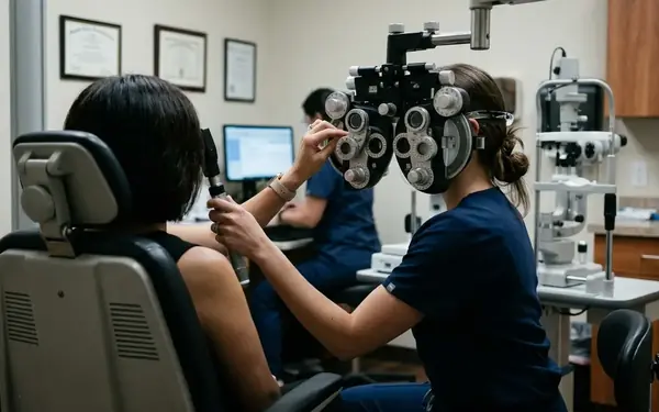
Diagnostic protocols previously relied heavily on non-contact tonometry for mass screening, but this approach was largely discarded as a standalone metric. Clinicians now mandate optical coherence tomography alongside traditional visual field testing.
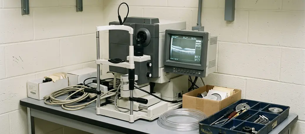
Based on longitudinal studies, optical coherence tomography can detect retinal nerve fiber layer thinning at the 70 to 80 micrometer threshold before visual field defects become apparent. This allows for intervention years before the patient notices a change in their functional vision. However, optical coherence tomography measurements can be confounded by severe cataracts or high axial myopia, which degrade image quality and alter normative database comparisons.
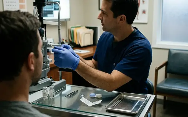
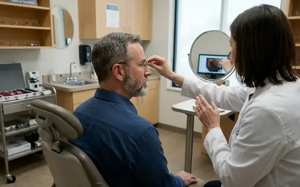
Critical Insight: A dilated eye exam and visual field testing are required for definitive diagnosis. The standard 'air puff' test alone is insufficient for ruling out optic nerve disease.
Preventative Strategies and Clinical Management
Proactive management begins with establishing a reliable baseline. High-risk demographics are advised to undergo dilated examinations every 12 to 24 months starting at age 40. Between visits, lifestyle modifications that support optimal vascular health, such as regular aerobic exercise and strict management of systemic diseases, can improve blood flow to the optic nerve.
When medical intervention is necessary, treatment algorithms dictate initiating therapy with topical prostaglandin analogs due to their once-daily dosing and efficacy in increasing uveoscleral outflow. Evaluating the efficacy of a new topical medication regimen requires a stabilization period of 4 to 6 weeks before assessing target pressure achievement. We only step up to beta-blockers or laser trabeculoplasty if target pressures remain unmet.
Selective laser trabeculoplasty is a proven secondary option for increasing aqueous outflow. However, the intraocular pressure-lowering efficacy of selective laser trabeculoplasty diminishes over time, often requiring a return to topical medications after a period of 36 to 60 months depending on the individual's trabecular meshwork response. For detailed protocols, practitioners often refer to the National Eye Institute clinical guidelines on glaucoma.
Recommendation: Adherence to prescribed topical medications is the single most important factor in preventing long-term vision loss in diagnosed patients.
While these diagnostic and treatment protocols represent current clinical standards, individual trabecular meshwork responses vary significantly, and no single intervention is guaranteed to halt progression in all patients.