Defining Over-the-Counter Cosmetic Contact Lenses
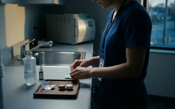
The distinction between a cosmetic accessory and a medical device blurs dangerously when a product sits directly on the eye. Over-the-counter decorative lenses are frequently marketed alongside makeup, false eyelashes, and costume accessories. This retail placement creates a false sense of security for consumers who view them as harmless fashion upgrades. Clinicians classify these products differently by evaluating their physical interaction with the tear film. They determine that any polymer resting on the corneal epithelium alters tear exchange, thus necessitating strict clinical oversight. This biological reality remains true regardless of whether the lens corrects visual acuity or simply changes iris color.
The tear film itself is a complex, three-layered structure essential for ocular health. When a contact lens is introduced, it splits this delicate film, altering evaporation rates and surface tension. Human corneas are highly individualized, complex anatomical structures that dictate how this interaction plays out. Corneal diameters typically range from 11.0 to 12.5 millimeters. Base curve radii require precise matching within an 8.3 to 9.0 millimeter range. When consumers bypass the prescription process, they completely ignore these fundamental anatomical metrics.
The FDA guidelines on decorative contact lenses classify all such products as medical devices to enforce this necessary oversight. Illegally distributed over-the-counter beauty products bypass regulatory scrutiny entirely. They place unverified, mass-produced materials directly onto a highly sensitive organ. A lens purchased at a novelty shop, a flea market, or an online boutique undergoes no clinical verification. The assumption that a non-corrective lens requires no medical supervision represents a fundamental misunderstanding of ocular physiology.
Physiological Consequences of Improper Lens Fitting
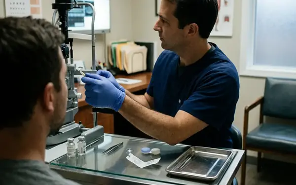
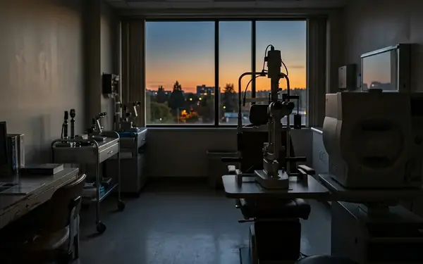
A single lens size cannot safely accommodate the natural variations in human corneal curvature. The concept of a universal fit in unregulated lenses is inherently dangerous. During a slit-lamp examination, practitioners assess lens fit by observing the conjunctival blood vessels. If blanching occurs at the lens edge, they determine the base curve is too steep and is actively restricting blood flow. A lens that fits too tightly acts like a suction cup. It traps cellular debris, metabolic waste, and environmental irritants beneath the surface. It also prevents fresh tears from washing over the cornea to deliver essential nutrients.
Conversely, a loose lens creates mechanical friction against the ocular surface with each blink. The human eye blinks thousands of times a day. This constant rubbing quickly leads to painful corneal abrasions, stripping away the protective outer layer of epithelial cells and exposing the sensitive nerve endings below.
Hidden Hypoxia Risks
Patients with naturally flatter corneas might not experience immediate mechanical friction from standard-sized unregulated lenses, though they remain fully susceptible to the hypoxic effects. Hypoxia, or severe oxygen deprivation, triggers a dangerous physiological cascade. Unregulated hydrogel lenses frequently exhibit oxygen transmissibility (Dk/t) values between 9 and 15. Modern clinical standards require Dk/t values exceeding 24 for daily wear to prevent hypoxia.
When the cornea starves for oxygen, the body attempts to compensate for the deficit. It responds by growing new, fragile blood vessels into the normally clear tissue—a condition known as corneal neovascularization that threatens long-term visual acuity. These new vessels can leak lipids and cause permanent scarring across the visual axis. The cheap materials used in unregulated lenses prioritize manufacturing cost over oxygen permeability, creating a hypoxic environment for the wearer.
Material Toxicity and Unregulated Manufacturing
The chemical composition of unregulated decorative lenses introduces severe risks independent of their physical fit. Legitimate manufacturers utilize complex, patented processes to encapsulate colorants safely. Illicit manufacturing environments frequently skip the crucial, expensive step of sealing dyes and tints within the lens matrix. Investigators initially attempted to test unregulated lenses using standard saline extraction methods, but discarded this approach upon realizing that unsealed pigments required simulated tear fluid conditions to accurately measure leaching rates. The complex chemistry of human tears interacts with these cheap pigments in unpredictable ways.
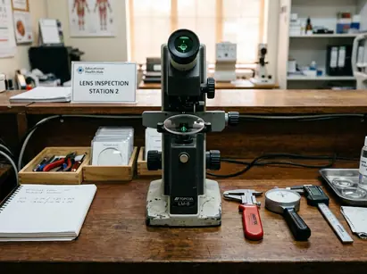
Pigment leaching tests reveal toxic exposure occurring within the first 3 to 4 hours of wear. The severity of pigment toxicity varies significantly based on the wearer's natural tear film composition, with lipid-heavy tears accelerating dye leaching from unsealed lens matrices. This chemical exposure causes toxic keratitis. The eye mounts an intense inflammatory response to the non-biocompatible pigments resting directly against the ocular surface. Symptoms include severe redness, photophobia, and a burning sensation that persists long after lens removal.
Microbiological Hazards in Packaging
Microbiological dangers compound the chemical risks. A person purchasing unregulated lenses from a beauty supply store can develop microbial keratitis within roughly 48 hours due to non-sterile blister pack fluid. Pseudomonas aeruginosa infections linked to non-sterile packaging can induce severe corneal ulceration within 24 to 48 hours. The lack of sterile manufacturing environments turns a simple cosmetic purchase into a direct vector for aggressive pathogens. These bacteria rapidly consume corneal tissue, utilizing the compromised epithelial surface as an entry point. Without immediate, aggressive medical intervention, such infections frequently result in permanent vision loss or the need for an emergency corneal transplant.
Clinical Protocols for Safe Cosmetic Lens Wear
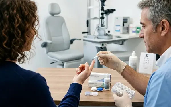
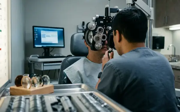
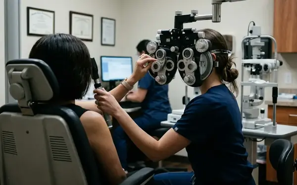
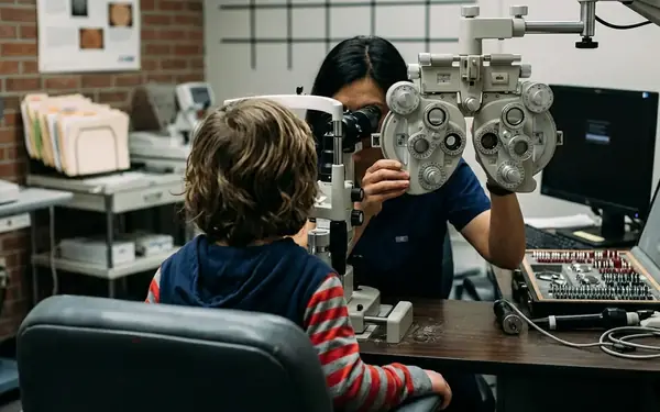
Changing eye color safely requires navigating authorized, prescription-based channels. The mandatory process begins with a comprehensive eye examination and a professional fitting. Practitioners evaluate the health of the ocular surface, assess tear production, and map the corneal topography before introducing any foreign material. The fitting protocol requires inserting a trial lens, mandating a 12 to 15-minute settling period, and then evaluating movement upon blinking to ensure adequate tear exchange before finalizing the prescription. This settling period allows the lens to equilibrate with the patient's body temperature and tear chemistry.
Clinical evaluations indicate that strict adherence to prescribed wear schedules prevents early-stage complications, though this observation is limited to patients using FDA-approved materials. Once a safe fit is confirmed, strict hygiene protocols dictate the daily routine. Initial wear schedules are strictly limited to 4 to 6 hours per day. This gradual introduction allows the cornea to adapt to the new physical barrier and helps the practitioner identify any early signs of intolerance.
Follow-up evaluations are scheduled 5 to 7 days after the initial fitting. During this visit, the clinician checks for microscopic signs of stress or inflammation that the patient cannot yet feel. Proper cleaning solutions and storage cases must replace the tap water or saliva that consumers sometimes dangerously substitute. A rigorous rub-and-rinse step is essential to remove protein deposits and environmental contaminants. Safe cosmetic wear is entirely possible. It simply demands the exact same medical rigor, patient education, and clinical oversight as corrective vision therapy.
The Long-Term Impact on Ocular Integrity
Severe corneal damage caused by unregulated lens wear often leaves irreversible scarring. The cornea relies on a delicate balance of fluid regulation to maintain its perfect transparency. Ophthalmologists evaluate long-term hypoxic damage by utilizing specular microscopy to map the endothelial layer, calculating cell density to decide if the cornea can safely sustain further contact lens wear. This specialized imaging provides a direct look at the cellular foundation of the eye, revealing damage that standard examinations might miss.
Healthy endothelial cell density ranges from 2,500 to 3,000 cells per square millimeter. These cells act as tiny, continuous pumps, constantly moving fluid out of the cornea to prevent swelling and clouding. Once these cells die off from chronic oxygen starvation or severe infection, they do not regenerate. The remaining cells must stretch and change shape to cover the gaps, reducing their overall efficiency. If the cell count drops too low, the cornea permanently swells, resulting in a painful condition known as bullous keratopathy.
The cornea is one of the only tissues in the human body that receives its oxygen directly from the air rather than the bloodstream. Atmospheric oxygen must dissolve into a tear film measuring roughly 3 to 5 micrometers thick to nourish the avascular cornea, making any unregulated, poorly fitted barrier a direct and immediate threat to cellular survival.