Navigating the First Signs of Near-Vision Strain
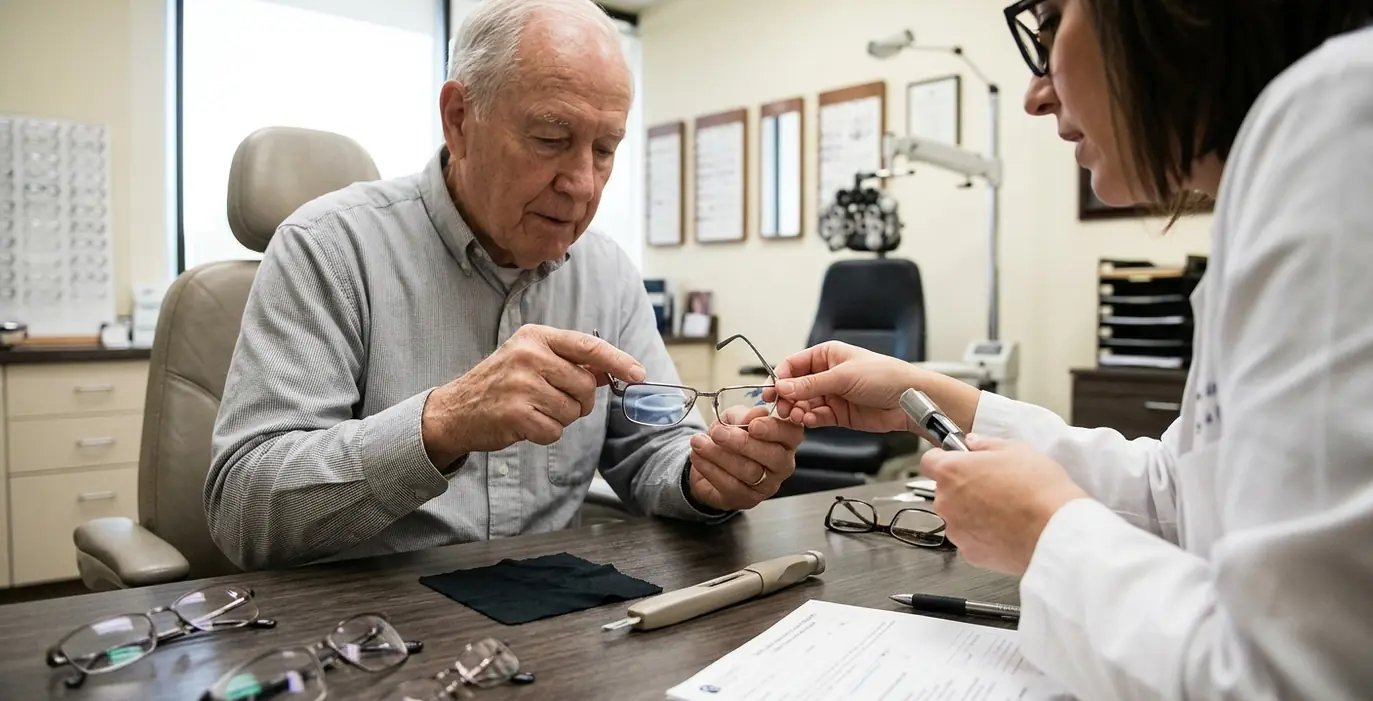
Why do perfectly measured progressive lenses often fail to provide immediate visual comfort for first-time wearers? This can stem from the complex relationship between optical physics and human neurology. Consider a recent case at a Broken Arrow optometry clinic involving a 45-year-old patient presenting with classic symptoms of presbyopia. The patient reported significant eye strain during extended computer work and increasing difficulty reading fine print on physical documents. These symptoms mark a standard physiological shift in the eye's crystalline lens, requiring a new approach to visual correction.
The crystalline lens naturally loses its elasticity over time, reducing the eye's ability to accommodate or change focus for near objects. This loss of flexibility forces the ciliary muscles to work harder. The resulting fatigue leads directly to the strain the patient experienced after hours of reviewing spreadsheets and digital reports.
The clinical team initially evaluated several corrective options to address these specific near-vision complaints. Standard lined bifocals offer a straightforward solution for near-vision deficits, providing a wide, clear reading area at the bottom of the lens. The patient's primary complaint involved focusing on a desktop monitor positioned at a viewing distance of 22 to 26 inches. Bifocals bypass this intermediate focal zone entirely. A bifocal wearer must lean forward awkwardly to use the reading segment or lean back to use the distance portion, leading to chronic neck and shoulder pain.
A progressive addition lens provides a continuous gradient of focusing power to accommodate multiple focal lengths simultaneously. The clinician prescribed a +1.25 diopter add power to support the patient's specific working distances, ensuring clear vision from the far wall of the office to the text on the screen and down to the paperwork on the desk.
The Architecture of Multi-Focal Lenses
Understanding the initial disorientation requires mapping the technical architecture of a progressive lens. The surface design incorporates distinct distance, intermediate, and near vision corridors blended into a single piece of material. The eye must navigate a central viewing channel while the brain actively suppresses the peripheral surface astigmatism inherent to the lens design. This peripheral distortion creates a visual sensation often described as a "swim effect"—a momentary feeling of spatial shifting during dynamic movement. The manufacturing process of blending multiple prescriptions inevitably pushes optical aberrations to the outer edges of the lens.
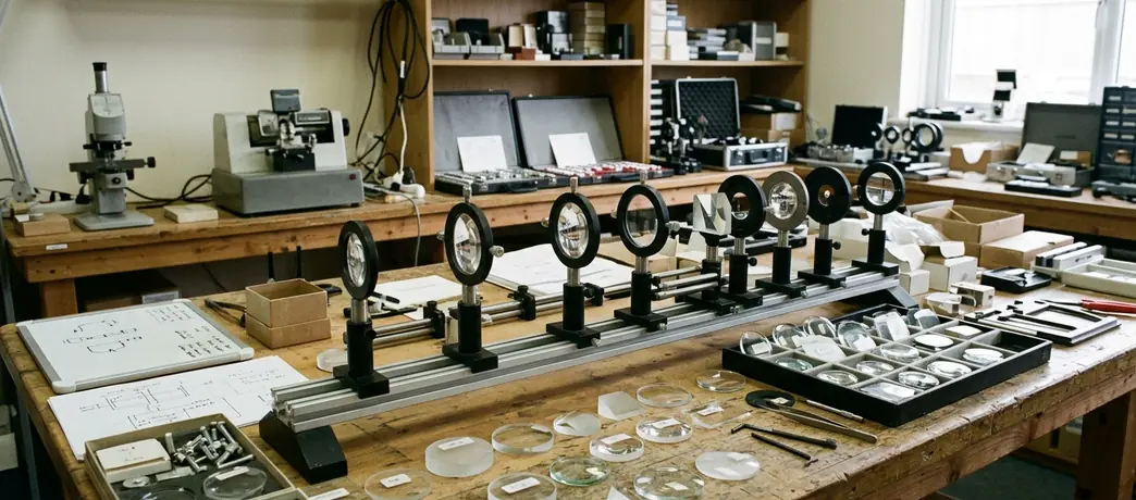
Clinical mapping of the intermediate corridor width typically reveals a functional viewing zone reported as roughly 3.8 to 4.5 millimeters. The brain must learn to keep the visual axis confined within this narrow channel during computer work. While clinical mapping provides a reliable baseline for lens design, individual neurological tolerance for peripheral distortion varies significantly among first-time wearers. The intensity of this peripheral distortion scales directly with the required add power. Prescriptions requiring an addition greater than +2.00 diopters will inherently present a narrower intermediate corridor regardless of the digital surfacing technology applied. The +1.25 diopter add power prescribed for this patient offered a relatively generous corridor, yet the initial adaptation still proved challenging.
The adaptation barrier also involves the disruption of the vestibulo-ocular reflex. This reflex coordinates eye and head movements to stabilize images on the retina, allowing a person to maintain focus on a stationary object while turning their head. Introducing a progressive lens alters the magnification and prismatic effects across the visual field. The eyes and inner ear temporarily send conflicting signals to the brain during head movement. Vestibulo-ocular reflex delay mapping in first-time progressive wearers indicates a processing lag reported as roughly 12 to 18 milliseconds. This microscopic delay manifests as mild dizziness or spatial disorientation until the neurological pathways recalibrate to the new optical environment.
Diagnosing Discomfort in the Exam Room
The patient returned for a follow-up examination reporting persistent nausea and specific difficulty navigating stairs. The optometrist systematically isolated the cause of the discomfort by evaluating the physical frame fit. A progressive lens demands exact alignment between the optical center and the patient's visual axis. The clinician first verified the optical center placement against the patient's natural pupillary distance and reading posture. Even a millimeter of misalignment can force the eye to look through the distorted peripheral zones, triggering the exact symptoms the patient described.
The physical frame fit plays a critical role in maximizing the usable field of view. The clinician measured the vertex distance, which dictates how far the back surface of the lens sits from the cornea. The initial measurement showed a vertex distance of 15 millimeters. A larger vertex distance effectively shrinks the wearer's field of view, similar to looking through a keyhole from a step back.
The evaluation also included the pantoscopic tilt, referring to the downward angle of the frame front relative to the vertical plane of the face. The frame exhibited a minimal pantoscopic tilt of 4 to 6 degrees. The resting posture on the bridge indicated that the transition corridor was sitting too low relative to the visual axis, forcing the patient to lift their chin unnaturally to access the intermediate and near zones.
Physical Calibration and Behavioral Retraining
The adjustment strategy focused on maximizing the usable field of view through precise physical calibration. By physically manipulating the nose pads, the clinician brought the lenses closer to the eyes. This intervention achieved a vertex distance reduction to 11 millimeters. Moving the lens closer to the eye dramatically expands the perceived width of the intermediate and reading corridors.
The clinician then angled the bottom rim inward, resulting in a pantoscopic tilt increase to 10 to 12 degrees. Pantoscopic tilt thresholds for intermediate corridor expansion demonstrate that these specific physical adjustments effectively widen the patient's perceived reading zone without altering the underlying lens prescription. The increased tilt aligns the reading zone naturally with the eye's downward rotation during near tasks.
Physical adjustments require complementary behavioral training to ensure long-term success. The clinician outlined a specific technique for lateral viewing: point your nose, not your eyes. This mantra trains the wearer to rotate their head toward the object of interest, keeping their visual axis aligned with the clear central corridor of the lens. Relying on peripheral eye movements, a common habit with single-vision lenses, forces the wearer's gaze directly into the areas of highest surface astigmatism.
Establishing a Structured Wear Schedule
To facilitate neuroadaptation, the clinician established a structured wear schedule. The protocol mandated an initial wear schedule of 3 to 5 hours daily, specifically targeting seated, stationary tasks. This controlled exposure allows the brain to process the new visual input without the added complexity of dynamic movement. The patient practiced shifting focus from a desktop monitor to a printed document, consciously aligning their head posture with the appropriate lens zone. Gradually increasing wear time builds neurological tolerance, preventing the sensory overload that often leads to early rejection of the lenses.
Tracking the Neurological Shift to Natural Sight
Evaluating the patient's progress required distinguishing between optical failure and normal neuroadaptation. The clinician reviewed the patient's progress at the two-week and four-week marks. The neuroadaptation window is reported as roughly 14 to 18 days, representing the critical period when conscious viewing effort transitions into a subconscious habit. During the initial days, the patient had to actively think about head positioning. By the two-week mark, the motor patterns began to automate.
During the two-week assessment, the patient demonstrated the ability to shift focus from a desktop monitor to a printed document without postural strain. The clinician assessed the patient's ability to perform dynamic tasks, such as walking down the hallway and navigating the clinic's staircase. The resolution of the swim effect during these dynamic activities indicated successful neurological filtering of the peripheral distortion. The brain had recalibrated the vestibulo-ocular reflex to account for the lens optics, eliminating the microscopic processing delay that caused the initial nausea.
The 28-day final clinical follow-up confirmed the long-term success of the fitting. The patient reported complete visual comfort throughout their workday and during evening activities. The initial nausea and spatial disorientation had entirely resolved. The combination of precise physical frame manipulation and strict adherence to the adaptation schedule yielded a highly functional visual outcome. The patient could now transition from driving to reading a text message with complete visual fluidity.
Committing to the Transition Timeline
The final consultation centered on establishing a clear baseline for future eyewear updates. Successful progressive lens wear requires both precise clinical fitting and patient commitment to the adaptation period. Initial discomfort represents a standard neurological response to new visual stimuli. The brain requires consistent exposure to build the necessary neural pathways for seamless, all-distance vision. Understanding this physiological reality helps patients manage their expectations and push through the challenging early days of wear.
The clinical data suggests a commitment period of roughly 21 to 30 days yields optimal results for first-time wearers. Rushing the process or reverting to old single-vision glasses disrupts the neuroadaptation cycle, forcing the brain to start over. The following table outlines the recommended progression through the adaptation phases, providing a roadmap for new wearers.
| Adaptation Phase | Timeframe | Recommended Activities | Visual Focus |
|---|---|---|---|
| Phase 1: Static | Days 1 to 3 | Reading, desktop computer work | Head-turning mechanics |
| Phase 2: Controlled Dynamic | Days 4 to 7 | Walking in familiar indoor spaces | Floor navigation, avoiding stairs |
Are you prepared to commit to the necessary neurological adaptation period to achieve seamless, all-distance vision, or does your daily routine demand the immediate, albeit limited, comfort of single-vision readers?