What a Small Pupil Can Hide During an Eye Exam
Declining pupil dilation can leave important areas of the peripheral retina inadequately examined. A retinal tear or diabetic retinal changes may develop before you notice a change in sight; glaucoma can also damage vision without obvious early symptoms. Missing these conditions can delay treatment and put vision at risk of permanent loss.
The limitation begins with the opening through which the optometrist looks. Examination light makes a naturally reactive pupil constrict, narrowing the view into the eye. Think of looking into a dark room through a small keyhole: you may see what sits directly ahead while missing what lies along the walls. An undilated examination can reveal central structures, including the macula and optic nerve, but obtaining a thorough peripheral view becomes more difficult.
Clear vision on the eye chart does not establish that the retina is healthy. Refraction measures which prescription brings an image into focus. It cannot, by itself, determine whether the optic nerve has damage or whether a tear lies near the retinal edge.
For patients arranging eye exams & care in Broken Arrow, that distinction matters when choosing an appointment. A full examination addresses eye health as well as the prescription for glasses or contact lenses. Dilation often supplies the access needed to examine the back of the eye thoroughly, although the examination plan should reflect your anatomy, symptoms, and clinical history.
The National Eye Institute explains the role of dilation in its comprehensive dilated eye exam guidelines.
How Diagnostic Drops Change Iris Muscle Activity
The iris regulates pupil size through opposing muscles. Its circular sphincter tightens to constrict the pupil in bright light. The radial dilator pulls outward to enlarge the opening. Diagnostic drops temporarily shift that balance so the examiner can direct light through a wider, more stable aperture.
Phenylephrine and Tropicamide Act Through Different Pathways
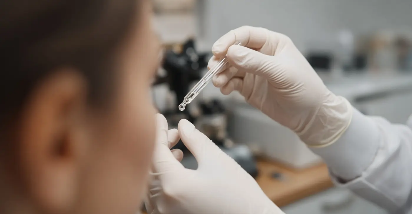
Phenylephrine stimulates the sympathetic pathway controlling the iris dilator muscle. Tropicamide blocks muscarinic receptors involved in parasympathetic muscle activity, relaxing the iris sphincter. Tropicamide also affects the ciliary muscle, which normally changes lens shape for near focusing. This reduction in accommodation is called cycloplegia.
These drug categories overlap: an antimuscarinic such as tropicamide produces both mydriasis, meaning pupil enlargement, and some cycloplegia. Phenylephrine primarily enlarges the pupil without directly relaxing accommodation. Combining agents can help maintain an adequate opening during examination, but the clinician selects the drugs and strength for the individual patient.
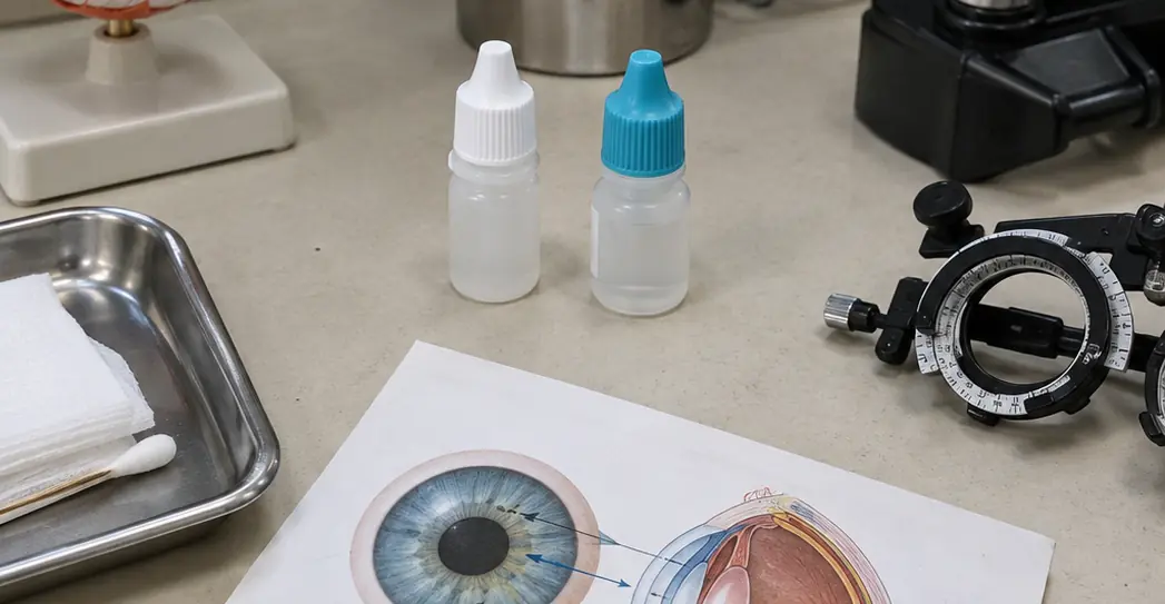
The Drainage Angle Needs Attention Before Dilation
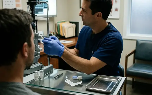
Before administering drops, the optometrist assesses the anterior chamber angle, where fluid drains between the cornea and iris. A slit-lamp estimate, often called a Van Herick assessment, helps identify potentially narrow angles. Gonioscopy provides a more direct examination when needed.
Narrow-Angle Precautions
In a susceptible eye, dilation can crowd peripheral iris tissue against the drainage pathway and trigger acute angle closure. This complication is rare but serious. A narrow-angle finding requires further assessment before routine dilation; the clinician may consider preventive treatment depending on the anatomy.
That anatomical check is an important boundary: a dilation regimen suitable for one patient may be unsafe for another.
What the Widened Pupil Reveals About the Retina and Optic Nerve
Once the drops take effect, the pupil resists the constriction that would otherwise interrupt examination under a bright beam. The optometrist can then use a slit lamp with a handheld lens to inspect central structures and a binocular indirect ophthalmoscope to examine more peripheral retina.
The examination follows anatomical landmarks, with a different question at each location:
- Optic nerve head: Does the central cup look enlarged or otherwise suspicious, and does the surrounding rim appear healthy? Cupping helps assess glaucoma risk, but its appearance alone cannot establish a diagnosis.
- Macula: Are there drusen, pigment changes, or other abnormalities in the retinal area responsible for detailed central vision?
- Peripheral retina: Is there lattice degeneration, a horseshoe-shaped tear, or another finding that needs closer assessment or treatment?
A finding also needs interpretation. Lattice degeneration, for example, does not automatically require treatment. Its significance depends on the surrounding retinal findings and the patient's symptoms.
Retinal Photography Adds Documentation, With Different Limits
Wide-field retinal photography records a broad view efficiently and preserves images for comparison over time. It can help patients understand a finding that would otherwise remain an abstract description.
A photograph and a direct examination provide different information. Stereoscopic examination supplies depth cues, while indirect ophthalmoscopy allows the clinician to change the viewing angle and investigate the far periphery. Eyelids, lashes, optical distortion, and image coverage can limit a photograph. A reassuring retinal image should not automatically replace an indicated dilated examination.
New flashes, a sudden increase in floaters, or a curtain-like shadow across vision warrant urgent eye assessment rather than waiting for a scheduled routine visit.
Planning for Glare and Near Blur After Your Broken Arrow Visit
Glare and focusing difficulty have separate causes. The enlarged pupil admits more light and cannot constrict normally in bright surroundings. Relaxation of the ciliary muscle makes near focusing harder—often most noticeable when reading a phone, a receipt, or dashboard controls.
Recovery takes several hours for many patients, with longer effects possible depending on the medication, its strength, and individual response. Iris pigmentation can influence drug uptake and persistence; lighter eyes may remain dilated longer, but eye color is an imperfect guide. Stronger cycloplegic agents, including those used for some children's focusing assessments, can cause more prolonged blur.
- Prepare before the appointment. Bring polarized sunglasses and arrange transport if you do not know how dilation affects your vision.
- Leave room in the day's schedule. Small print and screen-based tasks may remain uncomfortable while accommodation recovers.
- Check vision before driving. Sunglasses reduce glare but cannot restore focusing ability. Do not drive while blur or light sensitivity makes the road or instruments difficult to see.
Pain Needs Urgent Assessment
Severe eye pain, marked redness, worsening blur, halos around lights, headache, or nausea after dilation require urgent medical assessment. These symptoms can indicate acute angle closure and should not be dismissed as ordinary recovery.
Accept recommended dilation at your eye exam once the clinician has assessed its safety. Bring polarized sunglasses for the Oklahoma sun, and book a driver in advance if your response to the drops is unfamiliar.